ER: A Shift in the Night

each post gonna contain a bunch of cases i visited on ER or Clinic a week before

Wednesday, August 20, 2008

Clinical features and diagnosis of urinary tract infections in children


.
.
CLINICAL PRESENTATION


Younger children
In a meta-analysis of the diagnostic accuracy of the symptoms and signs of UTI in children younger than two years, the following symptoms and signs were the most helpful in identifying children with UTI


  • History of UTI (LR 2.3)

  • Temperature >40ºC (LR 3.2)

  • Suprapubic tenderness (LR 4.4)

  • Lack of circumcision (LR 2.8)

  • Temperature >39ºC for 48 hours in absence of another source for fever (LR 4.0)

  • Temperature <39ºc,>

Fever


  • Infants and children younger than 2 years can present with fever as the sole manifestation

  • The prevalence of UTI is greater among infants and young children with maximum temperatures 39ºC than in those with lesser degrees of fever

  • The presence of another potential source of fever (upper respiratory tract infection, acute otitis media, acute gastroenteritis) does not rule out the possibility of UTI

Other symptoms
Less common symptoms of UTI in infants include:


  • Conjugated hyperbilirubinemia (in those <28>
  • Irritability

  • Poor feeding

  • Failure to thrive

    Parental reporting of foul-smelling urine or the presence of gastrointestinal symptoms (vomiting, diarrhea, and poor feeding) does not correlate with UTI

Older children
Symptoms of UTI in older children may include:


  • Fever

  • Urinary symptoms (dysuria, urgency, frequency, incontinence, macroscopic hematuria)

  • Abdominal pain

The constellation of fever, chills, and flank pain is suggestive of pyelonephritis in older children

Occasionally, older children may present with:


  • Short stature

  • Poor weight gain

  • Hypertension secondary to renal scarring from unrecognized UTI earlier in childhood

  • Suprapubic tenderness and CVA tenderness may be present on examination of older children with UTI.

In a meta-analysis of the diagnostic accuracy of the symptoms and signs of UTI in verbal children, the following symptoms and signs were the most helpful in identifying children with UTI:


  • Abdominal pain (LR 6.3)

  • Back pain (LR 3.6)

  • Dysuria, frequency, or both (LR 2.2)

  • New onset urinary incontinence (LR 4.6)

CLINICAL EVALUATION

History

The history of the acute illness should include:


  • Height and duration of fever

  • Urinary symptoms (dysuria, frequency, urgency, incontinence)

  • Abdominal pain

  • Suprapubic discomfort

  • Back pain

  • Vomiting

  • Recent illnesses

  • Antibiotics administered

  • Sexual activity (if applicable)

Past medical history should include:


  • Chronic urinary symptoms (Incontinence, lack of proper stream, frequency, urgency)

  • Chronic constipation

  • Previous UTI

  • Vesicoureteral reflux (VUR)

  • Previous undiagnosed febrile illnesses

  • Family history of frequent UTI, VUR, and other genitourinary abnormalities

  • Antenatally diagnosed renal abnormality

  • Elevated blood pressure

  • Poor growth

Physical examination


Important aspects of the physical examination in the child with suspected UTI include:


  • Documentation of blood pressure and temperature.

  • Growth parameters (poor weight gain and/or failure to thrive may be an indication of chronic or recurrent UTI).

  • Abdominal examination for tenderness or mass (eg, enlarged bladder or enlarged kidney secondary to urinary obstruction).

  • Assessment of suprapubic and costovertebral tenderness.

  • Examination of the external genitalia for anatomic abnormalities.

  • Evaluation of the lower back for signs of occult myelodysplasia (e.g., midline pigmentation, lipoma, vascular lesion, sinus, tuft of hair), which may be associated with a neurogenic bladder.

  • Evaluation for other sources of fever.

LABORATORY EVALUATION


We recommend that urine samples be obtained for urinalysis (dipstick and microscopic examination) and culture in the following patients:


  • Girls and uncircumcised boys younger than two years with at least one risk factor for UTI (history of UTI, temperature >39ºC, fever without apparent source, ill appearance, suprapubic tenderness, fever >24 hours or nonblack race).

  • Circumcised boys younger than two years with suprapubic tenderness or at least two risk factors for UTI.

  • Girls and uncircumcised boys older than two years with any of the following urinary symptoms (abdominal pain, back pain, dysuria, frequency, high fever, or new-onset incontinence).

  • Circumcised boys older than two years with multiple urinary symptoms (abdominal pain, back pain, dysuria, frequency, high fever, or new-onset incontinence).
    Febrile infants and children with a abnormalities of the urinary tract, or family history of urinary tract disease.A different analysis highlighted the following risk factors:
    Age younger than 1 year
    White race
    Temperature 39ºC
    Fever for more than two days
    Absence of another source of fever on history or examination (ie, absence of upper respiratory infection, acute otitis media, gastroenteritis)

Urine culture is the gold standard for the diagnosis of UTI. We suggest that urine culture be performed in the following groups, even if the dipstick and microscopic analysis are negative:


  • Girls and uncircumcised boys younger than two years with at least one risk factor for UTI (history of UTI, temperature >39ºC, fever without apparent source, ill appearance, suprapubic tenderness, fever >24 hours or nonblack race).

  • Circumcised boys younger than two years with suprapubic tenderness or at least two risk factors for UTI.

  • Girls and uncircumcised boys older than two years with any of the following urinary or abdominal symptoms (abdominal pain, back pain, dysuria, frequency, high fever, or new-onset incontinence).

  • Circumcised boys older than two years with multiple urinary symptoms (abdominal pain, back pain, dysuria, frequency, high fever, or new-onset incontinence).
    Febrile infants and children with a abnormalities of the urinary tract, or family history of urinary tract disease.

  • Infants and children who will be presumptively treated with antibiotics.

Elevated peripheral white blood cell (WBC), erythrocyte sedimentation rate (ESR), and C-reactive protein (CRP) are indicators of an acute inflammatory process, but are not helpful in predicting UTI
In addition, though more sensitive than WBC and ESR, CRP does not reliably differentiate between cystitis and pyelonephritis (approximately 30 percent of children with a normal CRP have pyelonephritis)

Infants <1>

Clean catch sample — We define significant bacteriuria from a clean catch urine specimen in children as growth of 100,000 colony forming units (CFU)/mL of a single uropathogenic bacteria. This is the same as the standard definition for significant bacteriuria on clean catch specimens in adults, which is based upon studies from the 1950s.


Catheter sample — We define significant bacteriuria from catheterized specimens in children as growth of 50,000 CFU/mL of a single uropathogenic bacteria. (the American Academy of Pediatrics considers growth of 10,000 CFU/mL of a single pathogen as significant)
However, in a prospective study of febrile children <24>

Suprapubic sample — We define significant bacteriuria from suprapubic aspiration specimens as the growth of any uropathogenic bacteria (The American Academy of Pediatrics practice parameter considers the growth of any gram-negative uropathogen from a suprapubic specimen significant, but requires more than a few thousand CFU/mL for gram-positive pathogens)

Labels:

Tuesday, August 19, 2008

Acquired aplastic anemia in children and young adults


INTRODUCTION
  • Injury to or loss of pluripotent hematopoietic stem cells, in the absence of infiltrative disease of the bone marrow, is the major pathophysiologic characteristic of the disease
  • Acquired AA, characterized by pancytopenia and hypocellular bone marrow in the absence of abnormal infiltrates and without increased reticulum
  • The major constitutional or inherited causes of pancytopenia and AA in children include Fanconi anemia, dyskeratosis congenita, Shwachman- Diamond syndrome, and amegakaryocytic thrombocytopenia

ETIOLOGY AND INCIDENCE

  • The major identifiable etiologies are exposure to a wide variety of drugs and chemicals, ionizing radiation, and some viruses
  • AA may also rarely complicate orthotopic liver transplantation; AA in this setting has a very poor outcome except in those patients who develop the disorder in the background of fulminant hepatic
  • AA also has occurred in patients with other immune disorders such as systemic lupus erythematosus and, occasionally, in pregnancy

In one study in 213 children <17>

  • Radiation Exposure - Exposure to large doses of external radiation
  • Drugs - Numerous drugs, such as certain nonsteroidal antiinflammatory drugs (particularly phenylbutazone, which is now rarely used), chloramphenicol, gold, sulfonamides, certain of the antiepileptic agents, nifedipine, and cytotoxic drugs can be associated with
  • Other chemical agents –
  • Prolonged exposure to benzene is particularly notorious in this regard
  • Infectious agents - Patients with bacterial or viral diseases may experience pancytopenia.
  • This condition is usually transient and may relate to multiple factors, including the use of antibiotics and other medications
  • The best documented virus is parvovirus B19. However, pancytopenia can occur, particularly in immunocompromised patients.
  • Hepatitis - Hepatitis viruses and HIV can cause severe aplasia. The mechanism may involve T cell activation with release of cytokines. The responsible virus has not been identified; hepatitis A, B, C, and G do not appear to be involved
  • Pregnancy - Aplastic anemia associated with pregnancy frequently is self-limited, ending with delivery
  • Idiopathic - both immune system abnormalities as well as a genetic predisposition may play causative roles.

CLINICAL PRESENTATION AND DIAGNOSIS

The clinical presentation of AA is variable and includes symptoms and signs related to cytopenia in each of the three cell lineages:

  • Hemorrhagic manifestations secondary to thrombocytopenia
  • Fatigue, pallor, and cardiovascular complaints caused by progressive anemia.
  • Fever, mucosal ulcerations, and bacterial infections resulting from neutropenia

Diagnosis

A diagnosis of AA is suggested by the presence of pancytopenia with absolute reticulocytopenia, suggestive of bone marrow failure. (

The red blood cells usually are normocytic but occasionally may be macrocytic (mean cell volume >100)

The peripheral blood smear shows that the remaining elements, while reduced, are morphologically normal.

The differential diagnosis

  • Marrow replacement by fibrosis or tumor
  • severe megaloblastosis
  • paroxysmal nocturnal hemoglobinuria
  • myelodysplastic syndromes
  • Overwhelming infection caused by HIV or disorders associated with hemophagocytosis

The diagnosis of AA is established by bone marrow aspiration and biopsy. The characteristic findings include:

  • The marrow is profoundly hypocellular with a decrease in all elements; the marrow space is now composed of fat cells and marrow stroma
  • The residual hematopoietic cells are morphologically normal
  • Malignant infiltrates or fibrosis are absent
  • There is no megaloblastic hematopoiesis

Definitions of severity

  • Moderate aplastic anemia
    Bone marrow cellularity <50>6 weeks as documented by an absolute neutrophil count <1200/microl,>
  • Severe aplastic anemia
    A marrow biopsy showing less than 25 percent of normal cellularity or
    A bone marrow biopsy showing less than 50 percent normal cellularity in which fewer than 30 percent of the cells are hematopoietic and at least two of the following are present: absolute reticulocyte count <40,000/microl,>
  • Very severe aplastic anemia - The patient is considered to have very severe aplastic anemia (vSAA) if the ANC is <200/microl.>


Unless patients with SAA or vSAA are successfully treated, over 70 percent will be dead within one year
Spontaneous recovery is rare.

TREATMENT

  • Treatment includes withdrawal of offending agents (if any), supportive care, and some form of definitive therapy against the aplasia.
  • Blood and platelet transfusions should be used selectively in candidates for hematopoietic cell transplantation in order to avoid sensitization
  • Any blood products should be irradiated and CMV-negative, and should not be from family members
  • Patients with severe neutropenia affected with fever require immediate evaluation, blood culture, and treatment with broad spectrum antibiotics
  • Prophylactic antibiotics have no role

Severe AA— two major modalities for treatment of severe acquired AA are hematopoietic cell transplantation (HCT) and immunosuppressive therapy

For children with SAA or vSAA, including those with hepatitis- associated AA , the problems associated with HCT (eg, low frequency of matched sibling donors, graft failure, graft versus host disease) are outweighed by the advantages (eg, stable engraftment and hematopoiesis, low risk for clonal disorders such as myelodysplastic syndrome (MDS) or acute myeloid leukemia (AML)). Therefore, HCT from a matched sibling donor is the treatment of choice, with long-term disease-free survival rates approaching 90 percent

Immunosuppressive therapy - Only 20 to 25 percent of children with AA have an HLA-matched sibling. For the remaining children, intensive immunosuppressive therapy is the preferred option.

Initial regimens often consisted of antithymocyte globulin (ATG) alone, or cyclosporine alone, with response rates of about 50 percent
However, response rates of 75 to 80 percent currently are achieved by the use of more intensive regimens consisting of ATG, cyclosporine, corticosteroids, and hematopoietic growth factors

We currently use the following regimen:

  • Antithymocyte globulin (ATG) — 40 mg/kg per day for four days.
  • Cyclosporine (CSA) — 12 to 15 mg/kg per day in divided doses begun on day five to maintain a blood trough of 200 to 250 ng/mL.
  • Prednisone — 2 mg/kg (maximum 60 mg/day) for two weeks followed by a slow taper to prevent and control potential complications of serum sickness.
  • Recombinant human G-CSF or GM-CSF (eg, G-CSF at 5 microg/kg per day SQ for 90 days)

The response to therapy may be slow, with granulocyte recovery occurring first, followed by stabilization of hemoglobin and a decline in transfusion requirements. Platelet recovery may take months to years. Long-term survivors may show persistent thrombocytopenia, red blood cell macrocytosis, and elevated hemoglobin F concentrations

A variety of acute or delayed complications may be observed following immunosuppressive therapy in children with AA:

  • Serum sickness, occurring seven to 10 days after ATG therapy, is a common problem
  • Fever associated with the administration of ATG typically develops on the first day of treatment and lasts from one to six days.
  • Increased risk for opportunistic infections, particularly fungal infection
  • Cyclosporine has a variety of frequent side effects, including hypertension, nephrotoxicity, hirsutism, and gingival hypertrophy

The rate of relapse of aplasia increases over time, with reported values ranging from 9 percent at five years to 35 percent at fourteen years
Relapses typically respond to a second course of immunosuppressive therapy and, in responders, is not associated with a survival disadvantage
Nonresponders to repeat therapy have a worse outcome
Other than relapse, late complications following successful immunosuppressive therapy include evolution of clonal disease such as MDS and AML
The reported rate of these disorders has ranged from 9 percent at five years to 19 percent at ten years

Moderate AA –
For patients with moderate aplastic anemia (MAA), treatment recommendations are unclear
With progressive cytopenias, particularly severe neutropenia and/or transfusion dependence, treatment with HCT or immunosuppression should be considered
Further studies are needed for this variant of AA.

Labels: , ,

Sunday, August 17, 2008

Child Abuse

Friday, August 8, 2008

Supernumerary kidney

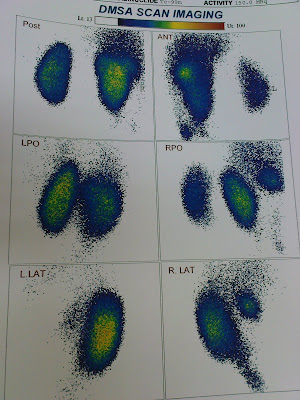
A 3.5 yo girl who admited with UTI month ago, was under treatment with prophilactic antibiotic (cefexim)

In US there was a hyronephrosis in left side and VUR in DRNC also


Here is her Renal Cortical Spect. Image; The right kidney showed with acceptable and homogeneous radiotrace uptake through out. There are two addition radiotracer uptakes in front of this kidney thath is better identified on 3D spect images The left kidney showed mildly diminished uptake through out. The differential renal function in the left kideny is 44% and right is 56% the buttom line was, Normal right renal cortical mass is noted. Mild dysfunction of left renal cortical mass is noted too. There is evidence of supernumerary kidney in the right side


Her physical exam and urin analysis was normal, so ABx therapy continued, and we asked for periodic U/A and U/C and anther US for next 3 month for following up the right kidney


Labels: , ,

Thursday, August 7, 2008

ITP

a 5yo girl with raccoon eyes (aka panda eyes) after a mild head truama; i though its a child abuse case at first sight but there was ecchimosis & petechia all over her body


Labels: , ,

Friday, August 1, 2008

Mitochondrial encephalopathy, lactic acidosis and cerebrovascular accident syndrome (MELAS)

Mitochondrial encephalopathy, lactic acidosis and cerebrovascular accident syndrome (MELAS) in PICU


Mitochondrial encephalopathy, lactic acidosis and cerebrovascular accident syndrome (MELAS) is, from the clinical point of view, one of the best studied mitochondrial multisistemic disorders. This disease has mainly been associated to the mitochondrial desoxyribonucleic acid (mtDNA) mutation A3243G located in the tRNALeu(UUR) gene. Although a relation between European haplogroups and the presence of the 3243 mutation has not been described

Labels: ,